ecg reading explained
Imagine your heart, a tireless engine, pumping blood through your veins with every beat. Now, picture a sudden, erratic rhythm, a chaotic dance that could disrupt the delicate balance of life itself. This is the reality for millions who experience heart arrhythmias, a condition often detected through an electrocardiogram (ECG) test.
An ECG is more than just a medical procedure; it’s a window into the intricate workings of your heart. By capturing the electrical signals that coordinate each heartbeat, an ECG can reveal hidden abnormalities, allowing for early diagnosis and timely intervention.
In this comprehensive guide, we’ll delve into the world of ECG readings, unraveling the mysteries behind the lines and curves that make up your unique heart rhythm. We’ll explore the basics of ECG interpretation, common abnormalities, and the significance of these findings for your overall heart health.
Understanding the Basics of an ECG
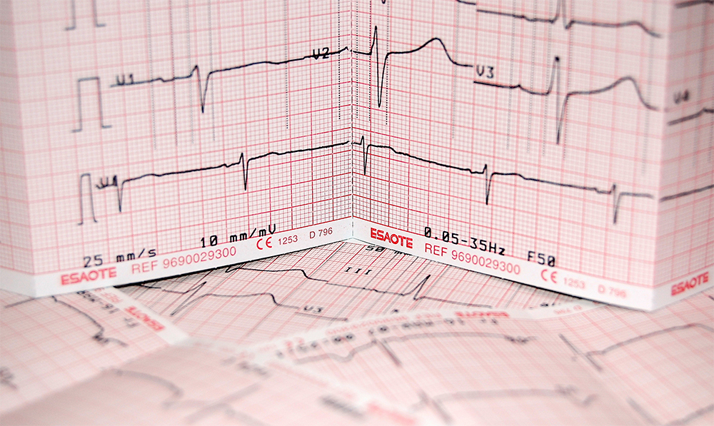
An electrocardiogram (ECG) is a non-invasive test that records the electrical activity of the heart. This electrical activity is essential for the heart to pump blood effectively throughout the body. By analyzing the ECG waveform, healthcare providers can identify potential heart problems and monitor heart health over time.
The P Wave The P wave represents the electrical activity associated with atrial depolarization. This is the initial phase of the heartbeat, where the electrical signal spreads across the atria, causing them to contract. A normal P wave is typically small, rounded, and upright. Abnormalities in the P wave can indicate various heart conditions, such as atrial enlargement or arrhythmias.
The QRS Complex The QRS complex is the most prominent part of the ECG waveform. It represents the electrical activity associated with ventricular depolarization. This is the phase where the electrical signal spreads through the ventricles, causing them to contract and pump blood out of the heart. The QRS complex is composed of three waves: the Q wave, the R wave, and the S wave. Abnormalities in the QRS complex can indicate various heart conditions, such as heart block or ventricular hypertrophy.
The T Wave The T wave represents the electrical activity associated with ventricular repolarization. This is the final phase of the heartbeat, where the ventricles relax and prepare for the next contraction. A normal T wave is typically upright and rounded. Abnormalities in the T wave can indicate various heart conditions, such as myocardial ischemia or electrolyte imbalances.
The U Wave The U wave is a small wave that sometimes follows the T wave. It is believed to represent the repolarization of the Purkinje fibers, which are specialized cells that conduct electrical signals through the ventricles. The U wave is not always visible on an ECG, and its significance is not fully understood.
The PR Interval The PR interval measures the time it takes for the electrical signal to travel from the atria to the ventricles. A normal PR interval is typically between 0.12 and 0.20 seconds. A prolonged PR interval can indicate a delay in the conduction of the electrical signal through the heart, which can be a sign of heart block.
A normal ECG, often referred to as a “sinus rhythm,” is characterized by a regular rhythm with consistent P, QRS, and T waves. Each heartbeat initiates in the heart’s natural pacemaker, the sinoatrial (SA) node, leading to a coordinated contraction of the heart chambers.
Sinus Bradycardia Sinus bradycardia is a condition where the heart rate is slower than normal, typically below 60 beats per minute. While it may be normal for some individuals, especially athletes, it can sometimes cause symptoms like fatigue, dizziness, or shortness of breath.
Sinus Tachycardia Sinus tachycardia is a condition where the heart rate is faster than normal, typically above 100 beats per minute. It can be a normal response to stress, exercise, or illness. However, persistent sinus tachycardia can be a sign of underlying heart problems.
Atrial Fibrillation Atrial fibrillation is a common heart rhythm disorder where the upper chambers of the heart (atria) beat irregularly and rapidly. This can lead to blood clots, stroke, and heart failure. An ECG can help diagnose atrial fibrillation by revealing irregular P waves or the absence of P waves altogether.
Atrial Flutter Atrial flutter is another heart rhythm disorder where the atria beat rapidly and regularly. However, unlike atrial fibrillation, the electrical signals in the atria are more organized. Atrial flutter can lead to rapid heart rates and can increase the risk of stroke.
Ventricular Tachycardia Ventricular tachycardia is a serious heart rhythm disorder where the ventricles beat rapidly. This can reduce the heart’s ability to pump blood effectively and can lead to sudden cardiac arrest. Ventricular tachycardia is often characterized by wide QRS complexes on an ECG.
Ventricular Fibrillation Ventricular fibrillation is a life-threatening heart rhythm disorder where the ventricles quiver chaotically, preventing the heart from pumping blood. Ventricular fibrillation is a medical emergency that requires immediate defibrillation.
Heart Block Heart block is a condition where the electrical signals that coordinate the heartbeat are delayed or blocked. This can lead to slow heart rates and other heart rhythm problems. Different types of heart block can be identified on an ECG, including first-degree, second-degree, and third-degree heart block.
ST-Segment Elevation Myocardial Infarction (STEMI) A STEMI is a type of heart attack where the blood flow to a part of the heart is completely blocked. This can cause significant damage to the heart muscle. An ECG can help diagnose a STEMI by revealing elevated ST segments in the affected leads.
Non-ST-Segment Elevation Myocardial Infarction (NSTEMI) An NSTEMI is a type of heart attack where the blood flow to a part of the heart is partially blocked. This can cause damage to the heart muscle, but the damage is usually less severe than a STEMI. An ECG can help diagnose an NSTEMI by revealing ST-segment depression or T-wave inversion in the affected leads.
Interpreting Your ECG Reading
While interpreting an ECG can be complex, understanding the basics can empower you to take charge of your heart health. By recognizing normal patterns and common abnormalities, you can work with your healthcare provider to make informed decisions about your well-being. Remember, an ECG is just one tool in the diagnostic toolbox. It’s essential to combine ECG results with other medical tests and a thorough physical exam for a complete picture of your heart health. If you have any concerns about your heart health or your ECG results, don’t hesitate to consult with a cardiologist. Early detection and treatment of heart conditions can significantly improve your quality of life.
And want to get some more out of your studying? Check out Paramedic Flash.
They have every flash card you could ever need to pass your medic class.
ECG strips, medications, and treatments, they’ve got you covered.
And if you want to help support the site, use my affiliate link here. It doesn’t cost anything more but it does help keep the site up and running.

Leave a comment